“Lenses and Image Formation”, an iBiology lecture by Prof. Daniel Fletcher (University of California, Berkeley). This lecture includes experimental demonstrations and provides a good review of the physics behind image formation by lenses.
- Light is described as being the visible (to humans) part of the electromagnetic spectrum. We learn about its properties such as wavelength and speed. Using a typical laser pointer as an example, Prof. Fletcher shows that its characteristics of wavelength, power, area and speed can be used to estimate its intensity, amplitude and frequency.
- The interaction of the laser pointer with a material such as glass is used to describe the principle of light-matter interaction that we know as refraction.
- Refraction and the focusing mechanism of Refractive lenses.
- Although light as an electromagnetic wave, “rays” are a convenient abstraction, and simplify the process of modeling optical systems with ray diagrams. This leads to an explanation of the ways in which images are formed by lenses.
Prof. Ron Vale’s iBiology lecture is titled “Microscope Imaging and Koehler illumination”. In this lecture, Prof. Vale shows how lenses are used in a microscope, building on the material in the previous lecture. He shows how lenses are used in image formation: to focus light from the specimen so that it can be viewed by our eyes through the oculars of a microscope or with a camera.
He also describes a method of illumination: how we use lenses to collect light from a light source to illuminate the sample, a technique called Koehler illumination. He also describes the conjugate planes of a microscope, a useful optical model for microscope users to conceptualize the operation of the versatile instruments that they use every day.
Dr. Jennifer Waters (Director of the Nikon Imaging Center at Harvard Medical School) provides a tutorial on how to align and optimize the illumination optics in the microscope for Koehler illumination.
Dr. Waters begins by showing “before and after” images. By showing images of the same sample taken on a misaligned system and then on a microscope that is properly setup with Koehler illumination, she makes a convincing case that this is a technique that all microscope users should learn.
Koehler illumination provides even illumination across the field of view and maximizes the contrast, while minimizing artifacts in the image. It is critical for obtaining high quality phase contrast and DIC images.
In this iBiology video, Prof. Ron Vale demonstrates how to set up Koehler Illumination on a microscope. This video provides a useful practical demonstration of the method described in the previous video.
Dr. Stephen Ross (Nikon) provides important information in this iBiology lecture about different classes of objectives and introduces the concepts of Point Spread Function (PSF) and Numerical Aperture (NA) as well as the tradeoffs between NA and Working Distance. He describes the use of immersion media to increase NA. Details are provided about the different types of aberration, such as spherical aberration and the use of a correction collar that may be used to correct it.
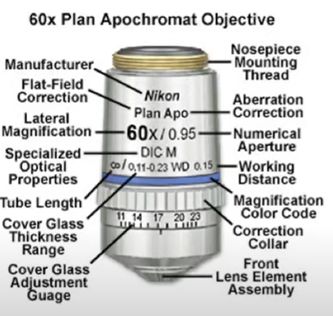
In this short iBiology video, Dr. Stephen Ross (Nikon) describes the key specifications that can be found by reading the information that is printed on a commercial microscope objective (shown below for reference).


